Introduction to Microscopes and Objective Lenses
Microscope objective lenses determine image resolution, magnification, and numerical aperture performance in microscopy systems. This video introduces the fundamentals and applications of microscope objective lenses.
Selecting the correct microscope objective lens is essential for achieving optimal imaging performance in microscopy systems. This video explains how magnification, numerical aperture, working distance, and application requirements influence objective lens selection.
Microscope Objective Lenses Explained
Types of Microscope Objective Lenses
- Scanning Objective Lens (4x): Used for locating and scanning specimens quickly with a wide field of view.
- Low Power Objective Lens (10x): Provides moderate magnification for general sample observation.
- High Power Objective Lens (40x): Designed for detailed imaging and higher-resolution observation.
- Oil Immersion Objective Lens (100x): Used for maximum resolution and high numerical aperture applications.
A microscope is a special optical device designed to magnify the image of an object. Depending on the type of microscope, it may project the image either onto a human eye or onto a recording or video device. As an example, consider the photographs of cells that can be found in a science textbook. These photographs have all been taken by a specialized microscope, and may be called micrographs.
While the simplest of microscopes is simply a magnifying glass with a single lens, compound microscopes used today are highly complex devices with a carefully designed series of lenses, filters, polarizers, beamsplitters, sensors, and perhaps even illumination sources. The exact combination of optical components used will depend on the application of the microscope; the wavelength of light with which it is intended to be used, and the resolution and magnification required in the final image.
The Components of a Basic Compound Microscope
A simple magnifier (magnifying glass), works when the object to be examined is situated within focal length of the magnifier lens, enabling larger virtual image is produced. This type of magnifier is very limited in both resolution and magnification. A compound microscope, on the other hand, uses a relay lens system instead of the single lens, and since each lens component can contribute magnifying power, the result is greatly increased capability.
Two major lens components—the objective lens and the ocular lens, or eyepiece—work together to project the image of the specimen onto a sensor. This may be the human eye or a digital sensor, depending on the microscope setup.
Since the objective is closest to the specimen being examined, it will relay a real image to the ocular lens. While doing so, it contributes a base magnification of anywhere from 4x (for a scanning objective lens, typically used to provide an overview of a sample) to 100x (for oil immersion objectives).
The ocular lens, located at the top of a standard microscope and close to the sensor (receiving eye) receives the real image from the ocular lens, magnifies the image received and relays a virtual image to the sensor. While most eyepieces magnify 10x, there are some which provide no magnification and others which magnify as much as 30x. The magnification power of the microscope can be calculated by multiplying the magnification power of the eyepiece, or ocular lens, by the magnification power of the objective lens. For example, an objective lens with a magnification of 10x used in combination with a standard eyepiece (magnification 10x) would project an image of the specimen magnified 100x.
Understanding Microscope Illumination
Since indirect backlight illumination is generally more effective than direct illumination, most microscopes do not include an internal light source. Instead, they rely on daylight or on background illumination such as a lightbulb. In brightfield illumination, also known as Koehler illumination, two convex lenses saturate the specimen with external light admitted from behind. These two lenses, the collector lens and condenser lens, work together to provide a bright, even, and constant light throughout the system: on the image plane as well as on the object plane. This system of illumination is used in many compound microscopes, including student microscopes and those found in many research labs.
Understanding Objective Lens
Microscope Objectives or Objective lenses are in many ways the heart of the microscope, and are typically mounted on a rotating nosepiece or turret to enable easy selection. Many microscopes will be equipped with a scanning objective (4x), a low power objective (10x), a high power objective (40x), and perhaps even an oil immersion objective lens.
Each microscope objective is itself a complex assembly of lenses, and besides contributing to the magnification, it is the objective lens which determines the resolution power of the microscope. An objective lens can also provide optical aberration corrections. A reflective objective, for instance, includes two mirrors within the assembly. These mirrors can focus laser light as well as provide chromatic corrections.
The Ocular Lens, or Eyepiece
The ocular lens, or eyepiece, is also an optical assembly rather than a single lens, but it is typically more simple than the objective. Often it is composed of two lenses: a field lens and an eye lens. The design of the ocular lens determines the field of view of the microscope, as well as contributing to the total magnification of the system.
Learn more about objective lenses by visiting our Deep Dive into Optics blog.
More on Microscope Objectives or Objective Lenses
A microscope objective is an important component of a microscopy or imaging system for a range of science research, biological, industrial, and general lab applications.. An objective lens determines the basic performance of an optical microscope or imaging systems and is designed for various performance needs and applications. It is located closest to the object and is an important component in imaging an object onto the human eye or an image sensor.
Objective lenses can be classified based on the objective construction, field of use, microscopy method, performance (optical aberration corrections), and magnification. Many microscope objective manufacturers offer a wide range of objective designs, which provide various degrees of optical aberration corrections for supporting different needs. Mirrors or reflective elements are used in objective lenses for the applications that requires chromatic aberration over board spectral ranges. Most traditional microscopy systems use refractive objectives such as achromatic objectives (the cheaper objectives) for laboratory microscope applications and plan apochromats (expensive objectives) for biological and science research microscope applications.
Objective Lens Specifications
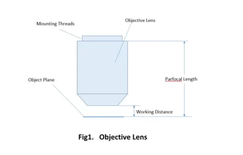
Important specifications are marked on the barrel of the objective, so students or researchers can easily identify the properties of an objective and determine the optical performance and working conditions for proper use. Figure 1 shows a diagram of an objective lens. A detailed discussion of the objection specifications is provided below.
- Magnification
Magnification is one important parameter. Magnification is usually denoted by an X next to a numeric value. Objectives are available in a range of magnifications from 2X to 200X.
- Magnification
- Numerical Aperture (NA)
NA is a critical value that indicates the light acceptance angle. It is commonly expressed as
- Numerical Aperture (NA)
NA = n × sinθ
where θ is the maximum 1/2 acceptance ray angle of the objective, and n is the index of refraction of the immersion medium. Figure 2 shows the ray angle θ of an infinity-corrected objective.
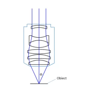
The value of NA determines the light gathering power and resolving power of the objective.
- Field of View
Field of View is the area of the object that can be imaged by a microscopy system. The size of the field of view is determined by the objective magnification or focal length of the tube lens for an infinite-corrected objective. In a camera system, the field of view of the objective is related to the sensor size.
- Field of View
- Optical Aberration Corrections
The optical aberration correction determines the optical performance of an objective lens and plays a central role in the image quality and measurement accuracy of imaging or microscopy systems. According to the degrees of the aberration corrections, objective lenses are generally classified into five basic types: Achromat, Plan Achromat, Plan Fluorite (Plan Semi-Apochromat), Plan Apochromat, and Super Apochromat.
- Optical Aberration Corrections
- Working Distance
Working distance is the free distance between the objective lens and the target.
- Working Distance
- Cover Glass Thickness
Many objectives are designed to be used with a cover glass. Using an incorrect coverslip thickness can greatly reduce the optical performance of a microscopy system.
- Cover Glass Thickness
- Parfocal Length
Parfocal distance is the distance between the objective mounting surface and the specimen when the image is in focus. Standard parfocal distances help microscope users switch between objective lenses without major refocusing.
- Parfocal Length
- Mounting Threads
For keeping the objective at the proper position, there are mounting threads on almost all objectives. Commonly used mounting threads include RMS, M25 x 0.75, M26X 0.706, M32 x 0.75.
- Mounting Threads
- Immersion Medium
Most objectives are designed to image specimens with air as the medium between the objective and the cover glass. However, for achieving higher working numerical apertures, some objectives are designed to image the specimen through another medium such as special oil with a refractive index of 1.51.
- Immersion Medium
How to Choose a Microscope Objective Lens
When selecting a microscope objective lens, important factors include:
- Magnification
- Numerical Aperture (NA)
- Working Distance
- Field of View
- Cover Glass Thickness
- Optical Aberration Correction
Choosing the correct objective lens helps improve image quality, resolution, and system performance.
Objective Lens Solutions
Objectives are complex multi-element lenses. For any given application, careful consideration of the optical parameters and specifications is necessary. In many cases, custom-designed objective assemblies provide the best-fit solution for meeting all the requirements of a specialized application. Custom parameters may include antireflection coatings, chromatic focus shift, working distance, image quality (MTF and spot size), lens mount, glass window thickness, and field of view, among others.
At Shanghai Optics, we design and manufacture custom objectives and imaging systems to support our customers’ needs in many industries, including medical, biomedical, machine version, scientific research, and metrology, etc. Taking the client’s budget and precision requirements into consideration, our experienced engineering team ensure that each design can be manufactured at a reasonable cost and the optical performance is being met based on fabrication, assembly, and alignment tolerance analysis.
Request a free quote from S.O. online, or contact a representative to learn more about our capabilities.
Frequently Asked Questions
What does a microscope objective lens do?
A microscope objective lens magnifies the specimen and determines image quality and resolution.
What is a scanning objective lens?
A scanning objective lens is typically a 4x lens used for locating samples quickly.
What is numerical aperture (NA)?
Numerical aperture measures the light-gathering ability and resolving power of an objective lens.
What is parfocal distance?
Parfocal distance is the distance between the objective and specimen when the image is focused.